Portable Venous Duplex Ultrasound Services
Rapid, Bedside Blood Clot & DVT Evaluations Brought to Your Facility
PERIPHERAL MOBILE IMAGING provides accurate venous ultrasound scans directly to physician practices, long-term care homes, correctional facilities, and homebound patients across Maryland, Washington DC, and Northern Virginia.
Personalized patient care
We deliver tailored health plans and services to meet your unique needs.
The results help healthcare providers determine the location and severity of venous reflux and guide treatment, compression therapy, or continued surveillance.
Portions of the examination may be performed while you are standing or in a
reverse Trendelenburg position to allow the veins to fill properly. Gentle compression or augmentation may be applied to the leg while Doppler ultrasound evaluates blood flow.
Our medical approach
Venous Insufficiency/ Venous Reflux
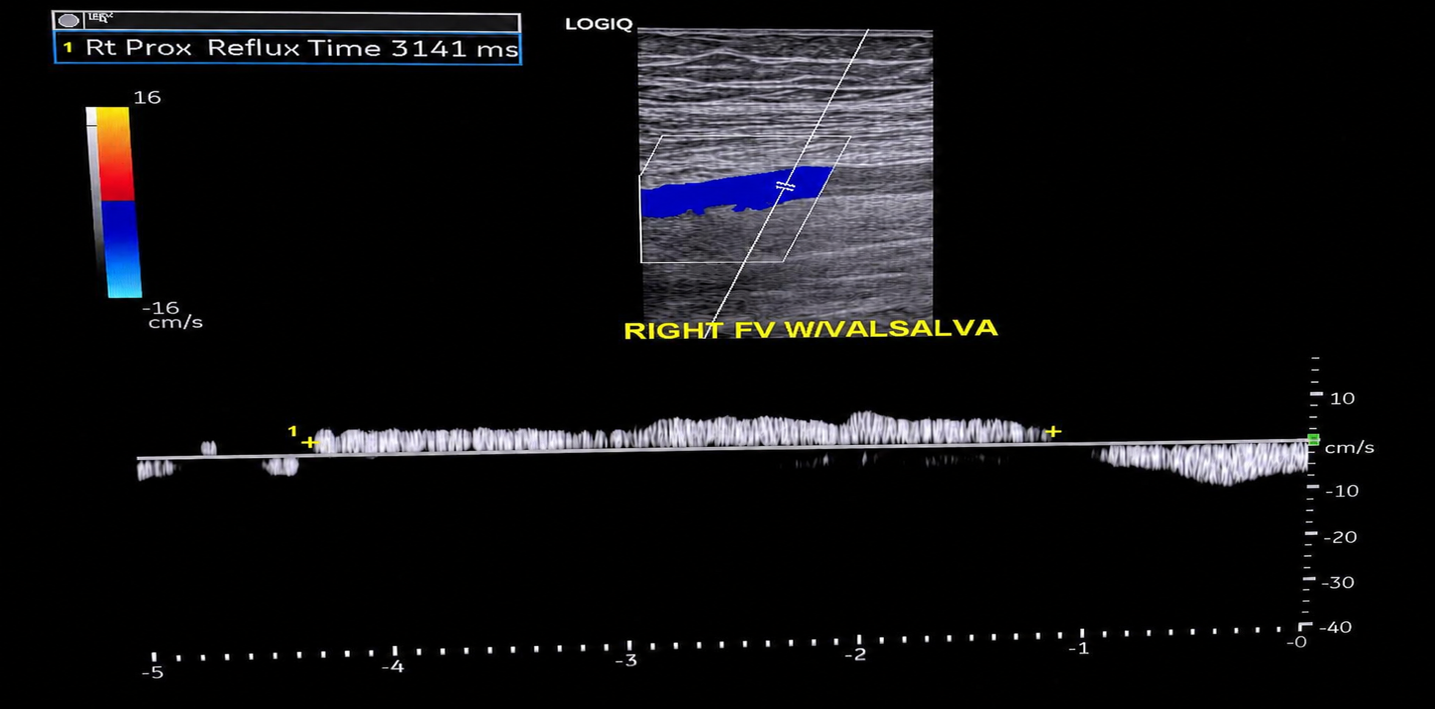
A venous insufficiency study, also called a venous reflux duplex ultrasound, is a safe, non-invasive examination that evaluates how well the veins in the legs return blood back toward the heart.
Healthy veins contain one-way valves that prevent blood from flowing backward. When these valves become weakened or damaged, blood may flow backward (venous reflux) and pool in the
legs. This condition is known as chronic venous insufficiency (CVI).
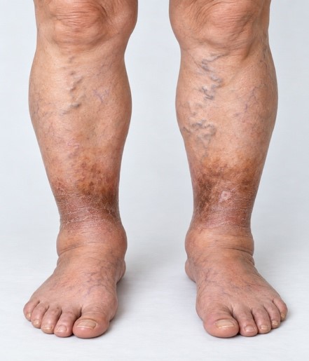
Venous insufficiency may contribute to symptoms such as leg swelling, aching or heaviness, varicose veins, skin discoloration, itching, and non-healing venous ulcers, particularly around the ankles.
During the examination, the technologist evaluates the deep and superficial venous systems, measures the direction and duration of blood flow, and identifies veins with abnormal reflux. The study may also help identify evidence of venous obstruction or previous deep vein thrombosis (DVT).
Long-Term Effects of DVT, a previous blood clot may leave behind residual thrombus, scar tissue, and damaged venous valves. When the valves no longer close properly, blood may flow backward (venous reflux) and pool in the leg. This can lead to post-thrombotic syndrome (PTS) or chronic venous insufficiency, causing persistent swelling, aching or heaviness, skin discoloration, and, in severe cases, venous ulcers. This is to show the impact of blot clot on the veins.

Key Benefits
Critical Prevention
Essential for early detection and immediate management of Deep Vein Thrombosis.
Safe & Non-Invasive
Safe for bedside use with no radiation, contrast agents, or special pre-procedure prep.
Full-Extremity Mapping
Evaluates deep and superficial vein networks in both arms and legs.
Common Indications for Venous Ultrasound
Ordering physicians and clinical care teams routinely request a mobile Venous Duplex scan when patients present with:
- Unilateral Swelling: Sudden or persistent edema in one leg, calf, or arm.
- Localized Pain or Tenderness: Unexplained leg cramping, throbbing, or deep soreness along vein pathways.
- Erythema & Warmth: Redness or localized skin warmth over a suspected vein thrombosis.
- High-Risk Immobility: Bedridden patients, recent surgical candidates, or long-term care residents with low mobility.
- Unexplained Shortness of Breath: Pulmonary embolism workup or suspected clot origin evaluation.
The Bedside Venous Examination
What to Expect During the On-Site Procedure
Positioning & Comfort
Compressibility Testing
Doppler Flow Assessment
Duration
Positioning & Comfort
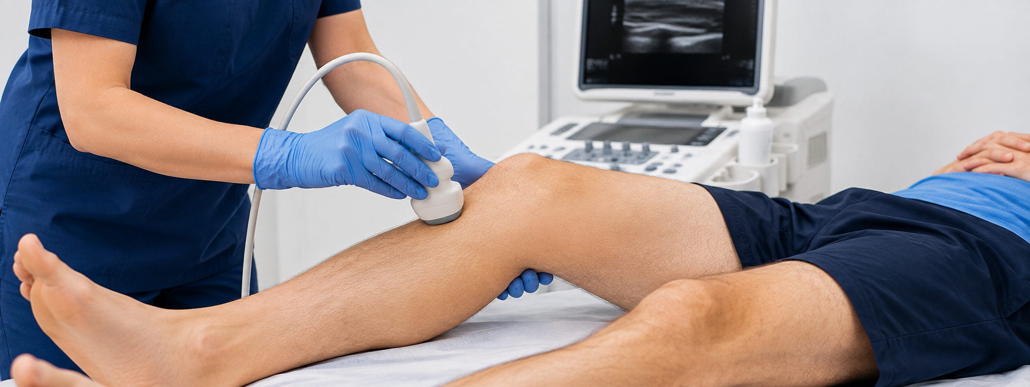
The patient is positioned comfortably on a bed or examination table, exposing the limb being evaluated.
Compressibility Testing
Our Registered Vascular Technologist applies gentle transducer pressure along key venous segments (e.g., femoral, popliteal, posterior tibial veins) to check vein compressibility.
Doppler Flow Assessment
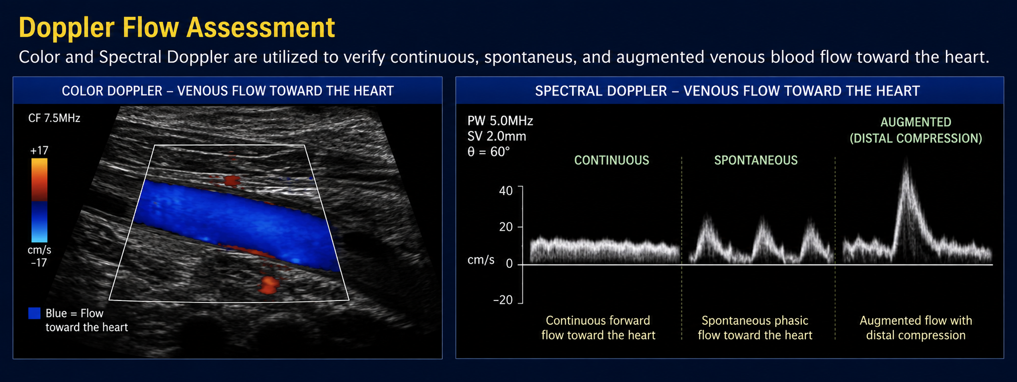
Color and Spectral Doppler are utilized to verify continuous, spontaneous, and augmented venous blood flow toward the heart.
Duration
The procedure typically takes between 30 and 45 minutes to complete on-site.
Turnaround & Reporting for Providers
Swift Diagnostic Delivery for Critical Decision-Making
Because DVT diagnosis often requires urgent medical intervention, Peripheral Mobile Imaging prioritizes rapid result transmission to your ordering team.
24–48 Hour Standard Turnaround
Comprehensive written preliminary and final diagnostic reports delivered straight to the referring clinic.
STAT Critical Call Protocol
Immediate phone/stat notification to the ordering physician if acute DVT or occlusive thrombus is detected.
Broad Insurance Coverage
Accepted by Medicare, Maryland Medicaid, CareFirst BCBS, UHC, Aetna, Humana, Cigna, TRICARE East, and major regional plans.
Creative
Bring On-Site DVT Testing Directly to Your Patients
Save high-risk or non-ambulatory patients the stress of emergency department visits and ambulance transport. Schedule a skilled, Certified Vascular Technologist portable scan in MD, DC, or Northern VA today.