Convenient, Bedside Aorta Imaging Brought to Your Facility
PERIPHERAL MOBILE IMAGING provides accurate aorta ultrasound studies at physician practices, long-term care facilities, correctional facilities, and homes across Maryland, Washington DC, and Northern Virginia.
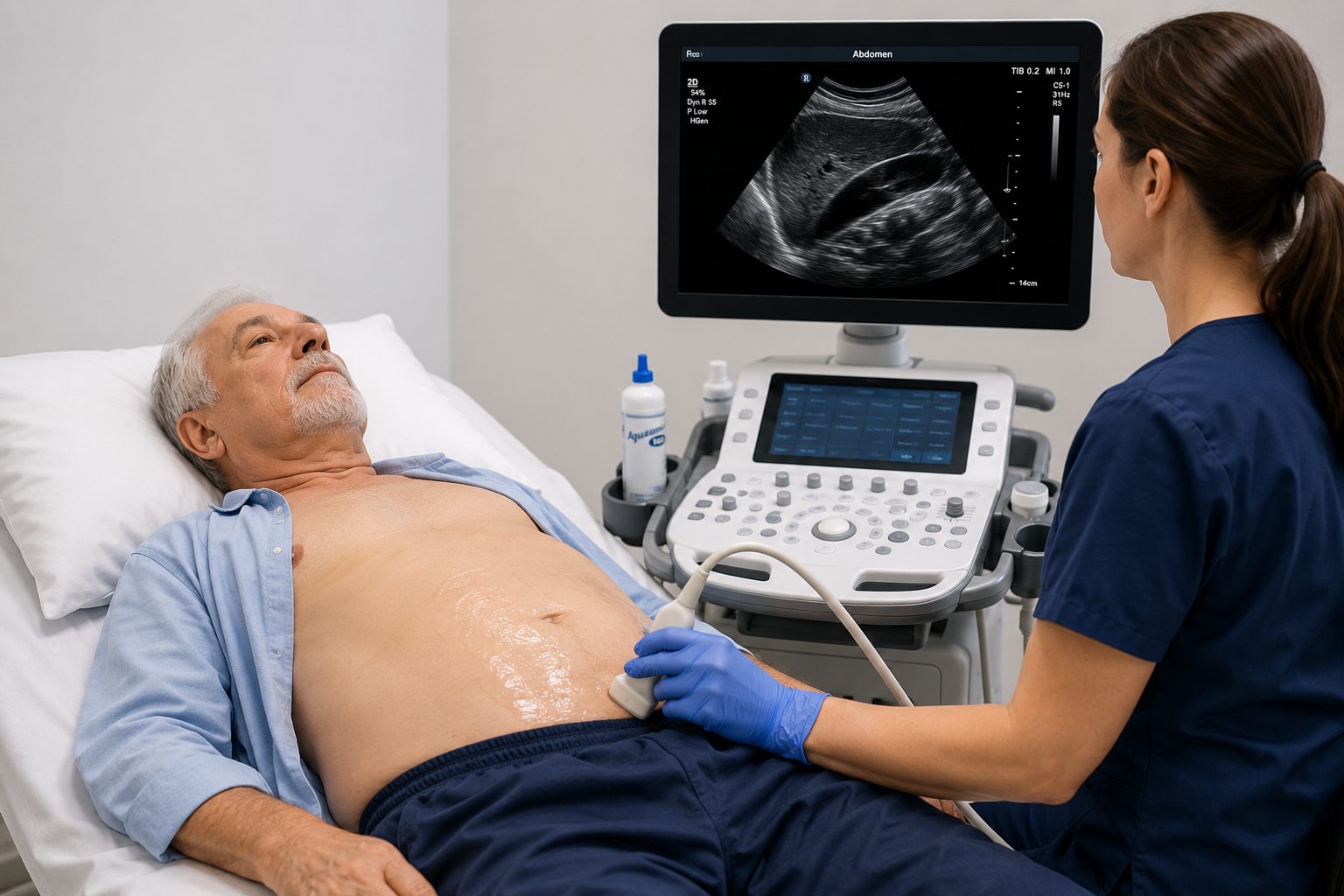
Our portable imaging helps evaluate aorta blood flow and vascular abnormalities conveniently at the patient’s bedside.
We provide detailed ultrasound assessment of aortic structure and blood flow to help identify aneurysms, narrowing, and other vascular abnormalities.
Our medical approach
Aorta Duplex Ultrasound
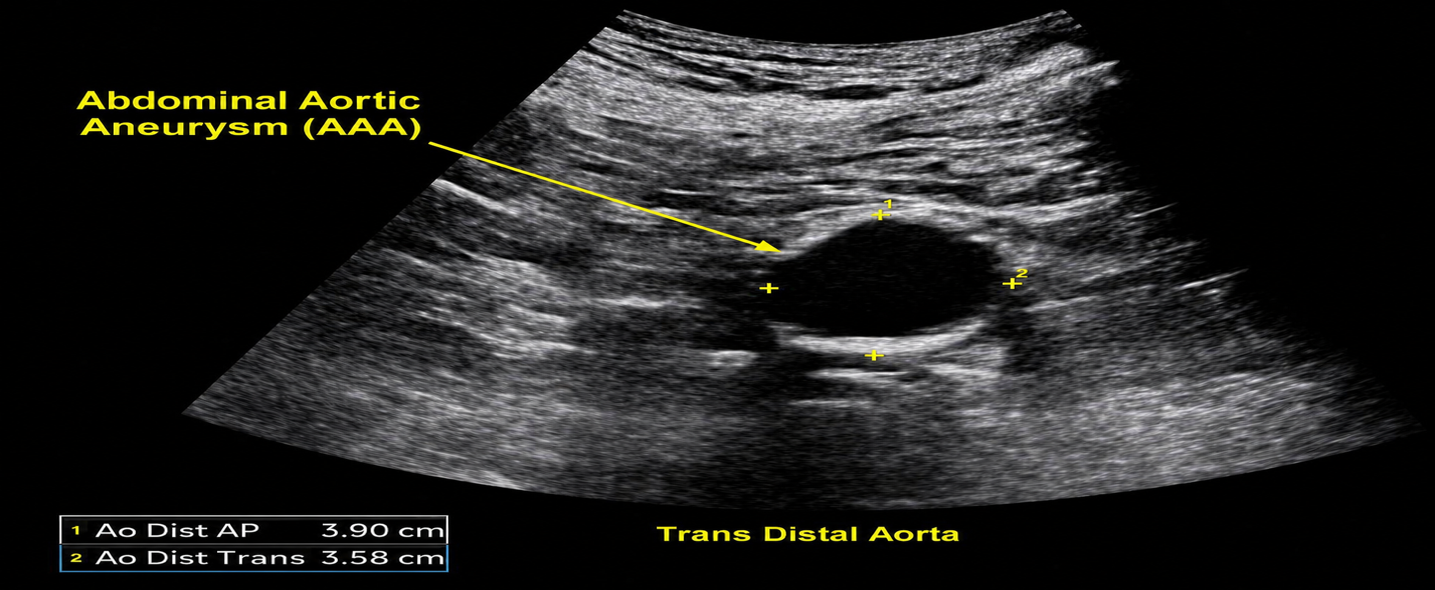
The abdominal aorta is the main artery carrying blood from the heart to the lower body. Ultrasound can evaluate its size and blood flow and help detect an abdominal aortic aneurysm (AAA), narrowing, or other vascular abnormalities.
Our medical approach
Mesenteric Duplex Ultrasound
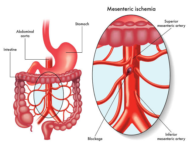
Evaluates the celiac artery and superior mesenteric artery (SMA), which supply blood to the digestive organs. Atherosclerotic plaque can cause narrowing (stenosis) or blockage, reducing blood flow to the intestines. Significant disease may be associated with abdominal pain after eating, food avoidance, and unexplained weight loss.
The examination can also help evaluate blood-flow changes associated with Median Arcuate Ligament Syndrome (MALS). MALS occurs when the median arcuate ligament of the diaphragm compresses the celiac artery, potentially restricting blood flow. Doppler ultrasound can demonstrate increased blood-flow velocities and changes with breathing that may suggest celiac artery compression. Further clinical evaluation and imaging may be necessary to confirm the diagnosis.
Our medical approach
Renal Artery Duplex Ultrasound
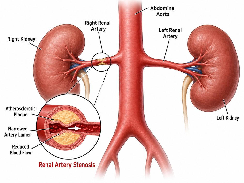
The renal arteries supply blood to the kidneys. Significant narrowing (renal artery stenosis) may contribute to difficult-to-control high blood pressure and, in some patients, impaired kidney function.
These examinations help identify vascular disease early and provide valuable information for diagnosis, treatment planning, and ongoing surveillance.
Key Benefits
Early Detection
Helps identify aortic aneurysms, narrowing, blockages, and other vascular abnormalities.
Safe & Non-Invasive
Uses ultrasound technology with no radiation or contrast agents.
Detailed Aortic Assessment
Evaluates aortic structure and blood flow to support accurate vascular evaluation.
Common Indications for Aortic Duplex Ultrasound
Ordering physicians and clinical care teams may request a mobile Aortic Duplex study when patients present with:
Known or Suspected Aneurysm: Evaluation of an enlarged or weakened aorta.
Abdominal or Back Pain: Unexplained symptoms that may require aortic evaluation.
Vascular Disease: Assessment of suspected aortic narrowing or blockage.
Abnormal Pulses: Changes in circulation that may indicate a vascular abnormality.
Aneurysm Monitoring: Follow-up imaging to monitor a known aortic aneurysm.
Patients should be NPO (Nothing by Mouth) before an abdominal vascular ultrasound. This means no food or drinks during the fasting period specified for your examination. Fasting helps reduce intestinal gas and improves visualization of the abdominal blood vessels. Please follow the specific preparation instructions provided for your appointment.
Aortic Imaging
Our technologist uses ultrasound to examine the abdominal aorta and assess its size, structure, and surrounding anatomy.
Doppler Flow Assessment
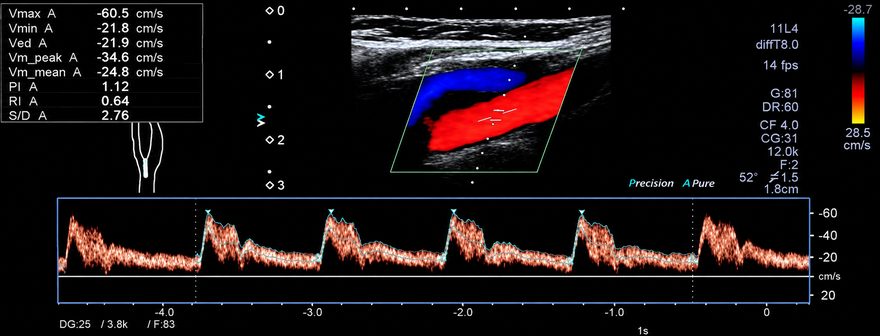
Color and Spectral Doppler are used to evaluate blood flow through the aorta and identify abnormalities.
Duration
An abdominal aorta duplex typically takes approximately 30–45 minutes, although examination times may vary depending on the complexity of the study.
Duration
A renal artery duplex is a more detailed examination and may take approximately 60 minutes or longer. Because the renal arteries are located deep within the abdomen and move with breathing, you may be asked to hold your breath briefly at different times during the examination. This helps stabilize the vessels and allows the technologist to obtain accurate images and Doppler blood-flow measurements.
Turnaround & Reporting for Providers
Swift Diagnostic Delivery for Critical Decision-Making
Because DVT diagnosis often requires urgent medical intervention, Peripheral Mobile Imaging prioritizes rapid result transmission to your ordering team.
Comprehensive preliminary and final diagnostic reports are delivered directly to the referring clinic.
STAT Critical Findings
Immediate notification to the ordering physician when significant aortic abnormalities, aneurysms, or critical blood flow issues are identified.
Broad Insurance Coverage
Accepted by Medicare, Maryland Medicaid, CareFirst BCBS, UHC, Aetna, Humana, Cigna, TRICARE East, and major regional plans.
Creative
Bring On-Site Abdominal Vasculatures to Your Patients
Provide convenient, bedside aorta evaluation for patients who may have difficulty traveling to an imaging center. Schedule a skilled, certified vascular technologist for abdominal vasculature in Maryland, DC, or Northern Virginia today.