Venous Duplex for Deep Vein Thrombosis (DVT)
Urgent Bedside Blood Clot Detection to Prevent Pulmonary Embolism
PERIPHERAL MOBILE IMAGING brings emergency and routine DVT evaluations directly to physician offices, long-term care facilities, correctional centers, and patient bedsides across Maryland, Washington DC, and Northern Virginia.
Early detection is important because a blood clot can potentially break loose and travel to the lungs, causing a pulmonary embolism (PE), which can be life-threatening.
Seek immediate medical attention for sudden shortness of breath, chest pain, coughing up blood, fainting, or other symptoms concerning for a pulmonary embolism.
Our medical approach
Deep Vein Thrombosis (DVT)
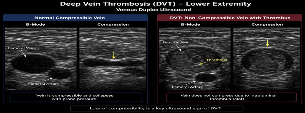
A venous duplex ultrasound is a safe, non-invasive examination used to evaluate blood flow through the veins and detect a deep vein thrombosis (DVT), a blood clot that forms within a deep vein, most commonly in the legs.
During the examination, the technologist will apply warm ultrasound gel to the extremity and gently use an ultrasound probe to compress and evaluate the veins, assessing for blood clots, abnormal blood flow, and venous obstruction. It’s an important part of identifying a DVT.
Common symptoms that may prompt a DVT examination include sudden leg or arm swelling, pain or tenderness, warmth, redness, or unexplained limb discomfort.
Key Benefits
Life-Saving Early Detection
Prevents fatal pulmonary embolisms by identifying acute or chronic deep vein clots early.
Non-Invasive & Radiation-Free
Safe for bedside testing with no needles, invasive probes, or contrast agents.
Immediate On-Site Results
Eliminates costly emergency room visits and uncomfortable patient transport.
Common indications for lower extremity artery blockages
Common indications for Deep Vein Thrombosis (DVT)
A venous duplex ultrasound may be ordered when a patient has:
- Sudden swelling of one leg or arm
- Calf, thigh, or arm pain and tenderness without a clear injury
- Warmth, redness, or skin discoloration
- Unexplained limb heaviness or tightness
- A noticeable difference in limb circumference
- Dilated superficial veins or unexplained extremity edema
- Suspected pulmonary embolism with concern for a clot source
- A previous DVT requiring follow-up
- Symptoms after surgery, hospitalization, prolonged bed rest, injury, or extended travel
- Symptoms associated with pregnancy, cancer, estrogen therapy, a central venous catheter, or a known clotting disorder
Emergency warning: Sudden shortness of breath, chest pain, coughing up blood, fainting, or a rapid heartbeat may indicate pulmonary embolism and require immediate emergency care.
Common PAD risk factors include:
- Smoking or history of tobacco use
- Diabetes
- High blood pressure (hypertension)
- High cholesterol
- Increasing age
- Obesity and physical inactivity
- Family history of vascular or cardiovascular disease
- Chronic kidney disease
- Established coronary artery or cerebrovascular disease
Early identification of PAD can help guide treatment and reduce the risk of worsening circulation, non-healing wounds, limb-threatening ischemia, and other cardiovascular complications.
The Bedside Scanning Process
What to Expect During the On-Site Procedure
Patient Preparation
Anatomical & Waveform Mapping
Velocity Analysis
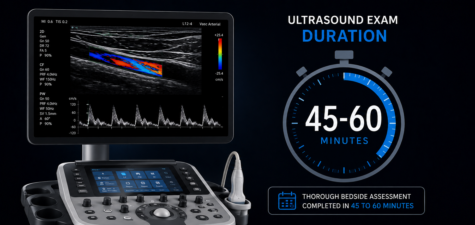
Duration
Patient Preparation
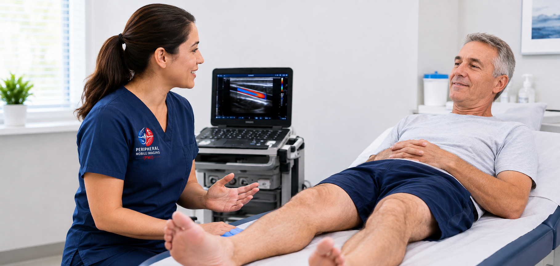
The patient rests in a comfortable supine position. The limbs to be examined are exposed while keeping the patient warm to avoid cold-induced vasoconstriction.
Anatomical & Waveform Mapping
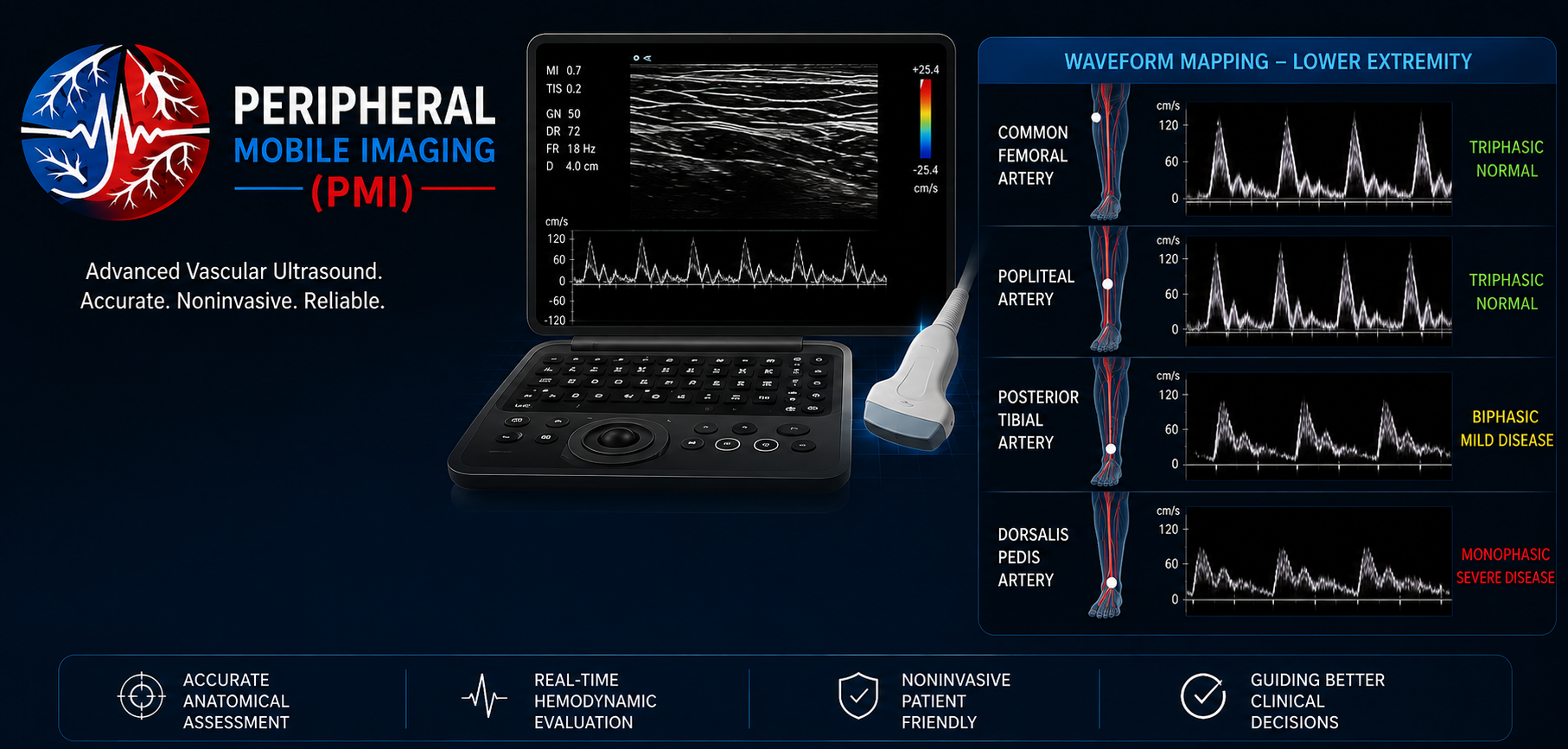
Our Registered Vascular Technologist glides an ultrasound transducer along the primary arterial pathways (e.g., femoral, popliteal, tibial, or brachial arteries).
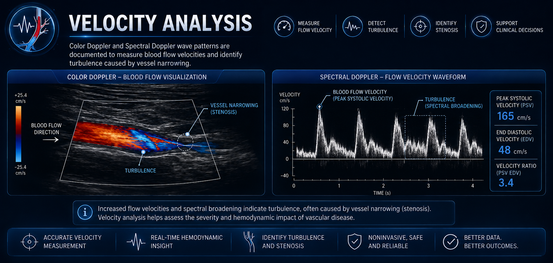
Velocity Analysis
Color Doppler and Spectral Doppler wave patterns are documented to measure blood flow velocities and identify turbulence caused by vessel narrowing.
Duration
Wear loose, comfortable clothing, preferably shorts, and loose-fitting undergarments to allow easy access to the groin area during the examination.
Empowering Brands
Reliable Preliminary Findings & Rapid Reports
Timely vascular evaluation is critical to saving limbs and guiding surgical or medical management. Peripheral Mobile Imaging ensures seamless data transmission to ordering providers.
24–48 Hour Standard Turnaround
Detailed written preliminary and final diagnostic reports delivered straight to the referring clinic.
Urgent Critical Findings Call
Immediate physician notification in cases of acute arterial occlusion or severe limb-threatening ischemia.
Insurance Coverage
Accepted by Medicare, Maryland Medicaid, CareFirst BCBS, UHC, Aetna, Humana, Cigna, TRICARE East, and major regional plans.
Creative

Schedule Portable DVT Diagnostic Scans Today
Deliver rapid blood clot evaluations to your patients across Maryland, Washington DC, or Northern Virginia.