Hemodialysis Access Evaluation

On-Site Dialysis Access Monitoring & Stenosis Prevention

PERIPHERAL MOBILE IMAGING delivers accurate AV fistula and graft evaluations directly to outpatient clinics, dialysis centers, skilled nursing facilities, and patient homes across Maryland, Washington DC, and Northern Virginia.

Our medical approach

Hemodialysis Access Evaluation

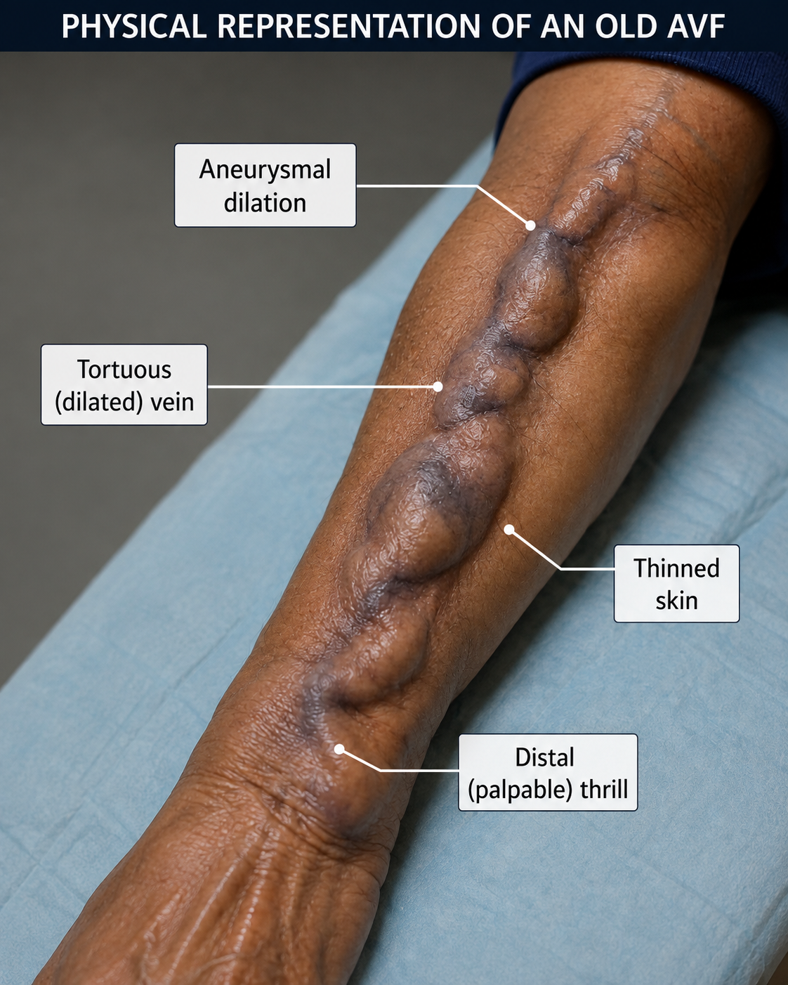
A Hemodialysis Access Evaluation is a specialized non-invasive vascular ultrasound scan used to assess the function, blood flow volume, and structural integrity of arteriovenous (AV) fistulas and synthetic AV grafts in end-stage renal disease (ESRD) patients. Over time, access sites are prone to developing juxta-anastomotic stenosis, neointimal hyperplasia, thrombosis, central vein stenosis, or aneurysmal degeneration. Routine portable ultrasound surveillance detects flow limitations early, preventing sudden access failure, inadequate dialysis clearance, and emergency hospitalizations.

Key Benefits

Access Lifespan Extension

Detects early stenosis to enable elective angioplasty or surgical intervention before complete access clotting.

Painless & Non-Invasive

Measures access volume flow (mL/min) and vessel diameters without needles or radiopaque contrast.

Bedside Care

Saves fragile dialysis patients unnecessary transportation stress and long waiting room visits.

Indications for Access Evaluation
Nephrologists, vascular surgeons, and dialysis care teams order portable hemodialysis access evaluations when patients present with:
  • High Venous Pressures: Persistent elevated arterial or venous pressure alarms during hemodialysis sessions.
  • Reduced Volume Flow: Decreased access flow rates (e.g., flow volume < 500–600 mL/min or a > 25% drop over time).
  • Physical Exam Abnormalities: Weak pulse, change in thrill/bruit (e.g., high-pitched pitch), localized swelling, or severe arm pain.
  • Maturity Check: Pre-dialysis evaluation to confirm AV fistula vein diameter (> 6 mm) and depth (< 6 mm) before first cannulation.
  • Ischemic Steal Syndrome: Coldness, numbness, weakness, or painful pallor in the hand or fingers during or between dialysis treatments.

The Bedside Scanning Process

What to Expect During the On-Site Procedure

Preparation & History

The technologist reviews the type of access (radiocephalic, brachiocephalic, brachiobruit/transposition, or PTFE graft), placement date, and recent dialysis machine metrics.

Comprehensive Anatomical Scan

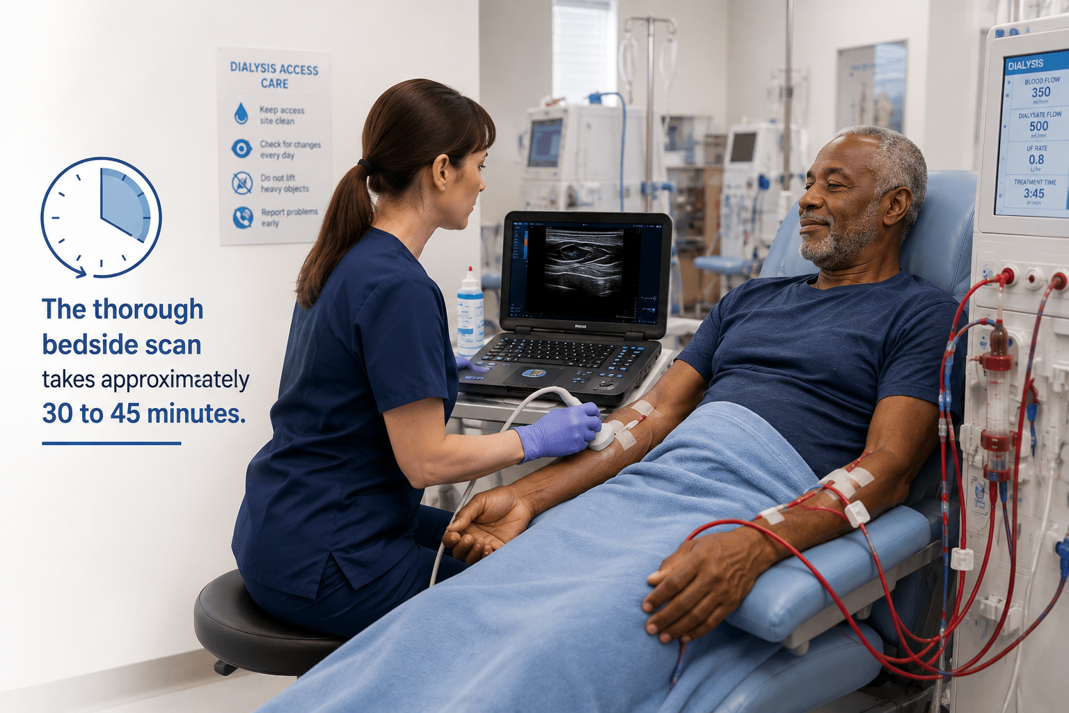
Our Registered Vascular Technologist applies gel along the feeding brachial/radial artery, the surgical anastomosis, the entire cannulation zone, and the outflow veins.

Volume Flow Calculations

Using High-Frequency Ultrasound and Color Spectral Doppler, the technologist measures vessel diameter and Time-Averaged Mean Velocity (TAMV) to calculate precise volume flow ($Q = \text{Area} \times \text{TAMV}$).

Duration

The thorough bedside scan takes approximately 30 to 45 minutes.

Empowering Brands

Fast Flow Metrics Delivered to Dialysis & Surgical Teams

Protecting vascular access lines requires rapid turnaround times so intervention can be scheduled without disrupting the patient’s dialysis schedule.

24–48 Hour Standard Turnaround

Detailed flow velocity, lumen diameter measurements, and written diagnostic reports sent directly to the referring nephrologist or surgeon.

STAT Critical Call Protocol

Immediate phone notification for acute access thrombosis, severe steal syndrome, or impending rupture of pseudoaneurysms.

Payer Acceptance

Accepted by Medicare, Maryland Medicaid, CareFirst BCBS, UHC, Aetna, Humana, Cigna, TRICARE East, and major regional plans.

Creative

Schedule Portable Dialysis Access Surveillance

Keep dialysis access lines functioning smoothly with  skilled, certified vascular technologists’ bedside diagnostics across MD, DC, and Northern VA.